FC13
CAMERA & FFA
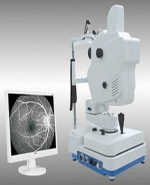
SPECIFICATIONS
Field Scope: 53 degree
The minimum checking pupil size: 3.3mm
Observing Light Source: Infrared light
Fixation: Internal Fixation and External Fixation
Color digital system: >12 Mega
Subsidiary function: two light dots Alignment.
Focus: Auto / Manual
Non-mydriatic Fundus camera in China
Easy operation, getting clear images within 10 secondes
Process the image anytime wanted, zooming, moving, sewing, measuring size.
Color fundus
Fundus fluorescein Angiography video and static flash shooting mode
Digital Color Fundus mode, Dynamic video Fundus fluorescein Angiography mode and static Flash Fundus fluorescein Angiography mode. |
Fundus Camera with Dicom 3.0 Interface in China, which can connect hospital’s HIS System, PACS System so that to achieve medical examination networked. |
Color Fundus Images, full digital process fundus Fundus fluorescein Angiography video and images. |
Process the images any time wanted, zooming, moving, paste, matching, measure and calculate focus size, editing and manage the cases. |
Small Pupil size fundus shooting, which is suitable for glaucoma. |
Internal fixation improves patient’s cooperation, improving the success rate greatly. |
Built-in digital acquisition system, the resolution can up to 1390 line. |
|